Article by m.l. chapagain and v.r. nerurkar - j. infect. dis. 2010

Human Polyomavirus JC (JCV) Infection of HumanB Lymphocytes: A Possible Mechanism for JCVTransmigration across the Blood-Brain Barrier Moti L. Chapagain and Vivek R. Nerurkar
Retrovirology Research Laboratory, Department of Tropical Medicine, Medical Microbiology, and Pharmacology, John A. Burns School of Medicine,University of Hawaii at Manoa, Honolulu, Hawaii (See the editorial commentary by Houff and Berger, on pages 181–183.)
It has been suggested that JC virus (JCV) might travel to the central nervous system in infected B cells.
Moreover, recent data suggest the presence of JCV in bone marrow plasma cells. However, the evidence for
infection and replication of JCV in B cells is unclear. To address this question, we infected Epstein-Barr virus–
transformed B cells with JCV and found that the viral genome decreased
11000-fold from days 0 to 20 after
infection, which concurred with the absence of viral early and late messenger RNA transcripts and proteins.
However, immunofluorescent images of B cells infected with fluorescein isothiocyanate–conjugated JCV dem-
onstrated that JCV enters the B cells, and DNase protection assay confirmed the presence of intact JCV virions
inside the B cells. Moreover, JCV-infected B cells were able to transmit infection to naive glial cells. These
data confirm that JCV nonproductively infects B cells and possibly uses them as a vehicle for transmigration
across the blood-brain barrier.

Progressive multifocal leukoencephalopathy (PML), a semination throughout the body and trafficking across subacute demyelinating disease of the central nervous the blood-brain barrier remains poorly understood.
system [1] results from the lytic infection of oligoden- A possible role of B cells in JCV transmigration across drocytes, the myelin-producing cells in the brain, with the blood-brain barrier has been suggested [8, 12, 14– human polyomavirus JC (JCV) [2–4]. JCV foci in a 18]. JCV-infected B cells were first detected in the spleen brain with PML are closely related to the blood vessels and bone marrow of 2 patients with PML [15] and [5], and JCV is presumably spread by the hematogenous were subsequently detected in the central nervous sys- route from the primary site of infection to secondary tem of a patient with PML [19]. Moreover, JCV DNA sites, such as kidneys, lymphoid tissues, and brain, to was reported to be associated with peripheral blood establish focal areas of infection or persistence [2, 6– lymphocytes in 89.5% and 38% of patients with PML 13]. However, the precise mechanism(s) of JCV dis- and patients with AIDS, respectively, with varying de-grees of immunodeficiency [7, 10]. Demonstration ofviral genome in the peripheral blood lymphocytes sug-gests the possibility of hematogenous spread of JCV.
Received 24 November 2009; accepted 14 January 2010; electronically published 15 June 2010.
Furthermore, JCV infection of human B cells in vitro Potential conflicts of interest: none reported.
was suggested [12, 18, 20], and it was argued that JCV Financial support: National Institute of Neurological Disorders and Stroke (grant R03NS060647), Research Centers in Minority Institutions Program (grants infection of Epstein-Barr virus (EBV)–transformed B G12RR003061 and P20RR011091), and Centers of Biomedical Research Excellence cells or B cell lines resulted in viral DNA replication (grant P20RR018727), National Center for Research Resources, National Institutesof Health, and Institutional Funds.
and production of infectious virions [18, 20]. However, Reprints or correspondence: Vivek R. Nerurkar, PhD, John A. Burns School of evidence for JCV replication in B cells was inconclusive, Medicine, University of Hawaii at Manoa, 651 Ilalo St, BSB 325AA, Honolulu, HI96813 ([email protected]).
because none of these studies quantitated the viral The Journal of Infectious Diseases
DNA, and messenger RNA (mRNA) transcripts were  2010 by the Infectious Diseases Society of America. All rights reserved.
rarely detected [18]. Moreover, infectious JCV virions in the B cells might be the residual virus inoculum used 184 • JID 2010:202 (15 July) • Chapagain and Nerurkar
to infect the B cells rather than de novo production of the 250, 1000, or 2500 HA units of JCV(Mad1) for 2 h at 37C, virus in the B cells.
were washed with phosphate-buffered saline (PBS) 3 times, and The possible role of B lymphocytes in JCV transmigration 2.5 ⫻ 10 cells were harvested at days 0 (2 h), 5, 10, across the blood-brain barrier came to light again in 2005 after 15, and 20 after infection, for DNA and RNA extraction.
the development of PML in patients with multiple sclerosis and Fluorescein isothiocyanate (FITC) labeling of JCV.
Crohn disease who were treated with a monoclonal antibody crose-purified JCV [31] was labeled with FITC using the Fluoro- natalizumab (Tysabri; Biogen Idec and Elan Pharmaceuticals) Tag FITC Conjugation Kit (Sigma) by modifying the published [21–23]. Natalizumab is a recombinant humanized monoclonal protocol [37, 38]. Briefly, 850 mL of the sucrose-purified virus antibody directed against the adhesion molecules a4b1 and containing 100 HAU/mL of JCV was dialyzed overnight with a4b7 integrins [24]. Because a4b1 integrin interacts with very labeling buffer (0.05 mol/L boric acid, 0.2 mol/L NaCl, pH 9.2), late antigen-4 (VLA-4) and this interaction is required for gen- and the volume was adjusted to 1 mL. The dialyzed virus (1 erating T and B cells from bone marrow progenitor cells in mL) was then transferred into a 2-mL tube containing a small adult mice [25], it is argued that a4-integrin blockade mobilizes stirrer, and 250 mL of FITC solution (1 mg/mL) in 0.1 mol/L JCV-infected pre-B cells from bone marrow into the circulation carbonate-bicarbonate buffer (pH 9.0) was added slowly drop and, thus, facilitates JCV dissemination and PML development by drop and incubated for 8 h at room temperature in the [26, 27]. Recently, 3 patients treated with efalizumab for pso- dark, with continuous gentle stirring. The FITC-labeled virus riasis also developed PML [28]. Efalizumab (Raptiva; Genen- was then dialyzed overnight with PBS (pH 7.2), was aliquoted, tech) is a humanized monoclonal antibody (IgG1) that binds and was stored at ⫺80C. As a control, 5 mg of bovine serum to the alpha chain (CD11a) of the leukocyte function associated albumin (BSA) in 1 mL of PBS was also conjugated with FITC, antigen (LFA-1) [29, 30]. LFA-1 is a member of the hetero- was aliquoted, and was stored at ⫺80C. Protein and FITC dimeric b2 integrin family, and it interacts with intercellular levels in the FITC-conjugated viral or BSA suspension were adhesion molecules (ICAM) expressed on antigen presenting determined according to the company's protocol (Sigma) by cells and endothelial cells and is necessary for T cell activation, measuring the absorbance at 280 nm and 495 nm, respectively.
T cell helper and B cell responses, natural killer cell cytotoxicity, The FITC-conjugated BSA was diluted to get the same absor- and antibody-dependent cytotoxicity [30]. Efalizumab can se- bance as FITC-conjugated JCV at 495 nm and was used as a lectively and reversibly block the activation, reactivation, and nonspecific fluorescence control.
trafficking of T cells [29, 30]. Natalizumab and efalizumab both Characterization of EBV-transformed B cell infection with
could create artificial cell-mediated immune deficiency in the One million B cells were incubated central nervous system by inhibiting the migration of lympho- with 1000 HAU of FITC-conjugated JCV (FITC-JCV) or FITC- cytes across the blood-brain barrier and, thus, may facilitate conjugated BSA (FITC-BSA) containing the same amount of the development of PML.
fluorescence in 500 mL of RPMI-1640 for 2 h. The B cells were To better understand the possible role of B cells in JCV washed 3 times with PBS, and the pellet was dissolved in 1 mL transmigration across the blood-brain barrier, we studied the of PBS. Forty microliters of cell suspension containing ∼40,000 kinetics of JCV(Mad1) infection of B cells in vitro. Our data B cells were transferred into each well of a multiwell slide, were confirm that JCV nonproductively infects B cells. However, JCV air dried, and were fixed with 4% paraformaldehyde for 10 virions persist in B cells, and B cells can transmit infectious min. Cells were then washed with PBS and permeabilized with virions to naive primary human fetal glial (PHFG) cells, sug- 0.4% Triton-X 100 for 5 min. The permeabilized cells were gesting that B cells can act as a vehicle for JCV transmigration blocked with 5% BSA for 1 h and were incubated with anti- across the blood-brain barrier.
rabbit protein disulphide isomerase (1:500), to visualize theplasma membrane, and were washed 3 times with PBS. The MATERIALS AND METHODS
cells were further incubated with anti-rabbit secondary anti- Virus and cell cultures.
JCV(Mad1) was propagated in PHFG body conjugated with Alexa-594 and were mounted with Vec- cells and was purified and quantitated by the HA assay and tashield mounting medium with DAPI (Vector Laboratories).
real-time polymerase chain reaction (PCR) [31–34]. EBV-trans- Fluorescent cells were examined using a Axiocam MRm camera formed primary B cells were provided by Dr Allison Imrie, mounted on a Zeiss Axiovert 200 microscope, equipped with University of Hawaii, and were cultured in RPMI-1640 sup- appropriate fluorescence filters and objectives as described else- plemented with 10% fetal bovine serum, penicillin (100 U/mL), streptomycin (100 mg/mL), and 2 mmol/L L-glutamate at 37C, One million EBV-transformed B cells were incubated with with 5% CO as described elsewhere [35, 36].
125, 250, 500, or 1000 HAU of FITC-JCV for 2 h and were JCV infection of EBV-transformed B cells.
washed with PBS. Cells were incubated with antibody against 2.5 ⫻ 10 cells) in suspension were infected with CD19 conjugated with PE (CD19-PE) and were subjected to JCV Infection of Human B Lymphocytes • JID 2010:202 (15 July) • 185


flow cytometry in Guava EasyCyte Plus platform. BSA labeledwith FITC containing the same amount of fluorescence wasused as a control.
DNA and RNA extraction.
Uninfected and JCV-infected B cells in T25 flasks were washed with PBS, were counted, andan aliquot of 0.25 million cells in duplicate were either frozenfor DNA extraction or lysed with 350 mL of RLT plus bufferfor RNA extraction (Qiagen) and stored at ⫺80C. RNA wasisolated using protocols reported elsewhere [32]. Additionally,100 mL of DNA was extracted using the Qiagen QIAprep SpinMiniprep Kit from each 0.25 million cells harvested at differenttime points, according to the manufacturer's protocol.
Quantitation of viral DNA and mRNA transcripts and re-
verse transcription PCR.
Two microliters of template DNA or complementary DNA were amplified and quantitated in theBio-Rad's iCycler iQ Multicolor Real-Time PCR Detection Sys-tem with use of primers, probes, and protocols described else-where [39]. Copies of JCV TAg or VP-1 genomes or mRNAtranscripts in experimental samples were calculated from thestandard curve and expressed as copies of viral genome per250,000 B cells or mRNA transcripts per microgram of totalRNA [32, 39].
JCV infection of B lymphocytes is nonproductive.
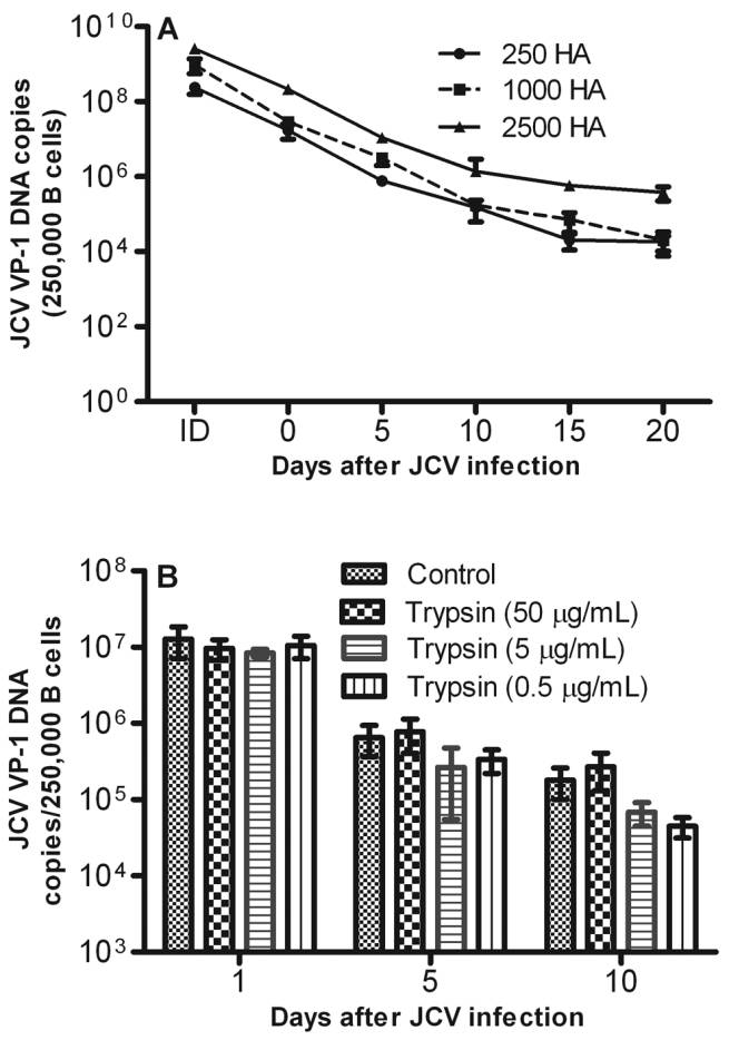
1 genome copies recovered from 2.5 ⫻ 10 infected B cells de- creased 11000-fold from day 0 to day 20 after infection (Figure1A) and neither early (TAg) nor late (VP-1) mRNA transcripts Figure 1.
JC virus (JCV) infection of B cells is nonproductive. Epstein- were detected (data not shown), suggesting that JCV infection Barr virus–transformed B cells ( 2.5 ⫻ 10 ) were infected with 250, 1000, or 2500 HA units of JCV(Mad1) for 2 h, and aliquots of of B cells is nonproductive. Our quantitative PCR and quan- were harvested at days 0 (2 h), 5, 10, 15, and 20 after infection. A, DNA titative reverse transcription PCR assays were sensitive and con- was extracted from 2.5 ⫻ 10 cells, and VP-1 DNA was amplified and sistently detected as low as 10–100 copies of JCV DNA and quantitated by quantitative polymerase chain reaction. VP-1 DNA genome mRNA transcripts, respectively [31]. Because JCV infection of copies decreased exponentially from days 0 to 20 after infection. B, Trypsin EBV-transformed B cells did not result in viral genome repli- treatment had no effect on further reducing the JCV genome copies at cation or transcripts expression, it is possible that JCV might any time point. JCV VP-1 genome copies recovered from 25, 100, or 250HAU of JCV equivalent used to infect 250,000 B lymphocytes was shown have remained associated with the B cells by attaching to the for comparison. ID, infection dose.
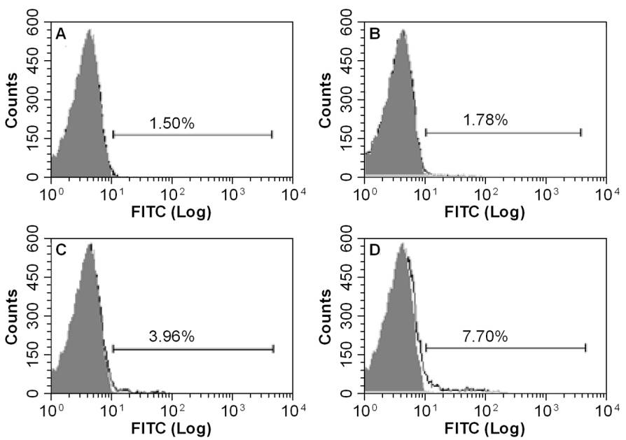
cell membrane and might not have entered the B cells. To testthis hypothesis, 2.5 ⫻ 10 B cells were inoculated with 250 HAU of JCV and incubated for 2 h at 37C. After incubation, the cells were incubated with 125, 250, 500, or 1000 HAU of JCV cells were washed twice with PBS and trypsinized for 10 min per million B cells, ∼1.5%, 1.7%, 3.9%, or 7.7% of B cells, with 0.5, 5, or 50 mg/mL of trypsin or with PBS alone, followed respectively, were infected with JCV (Figure 2A–2D).
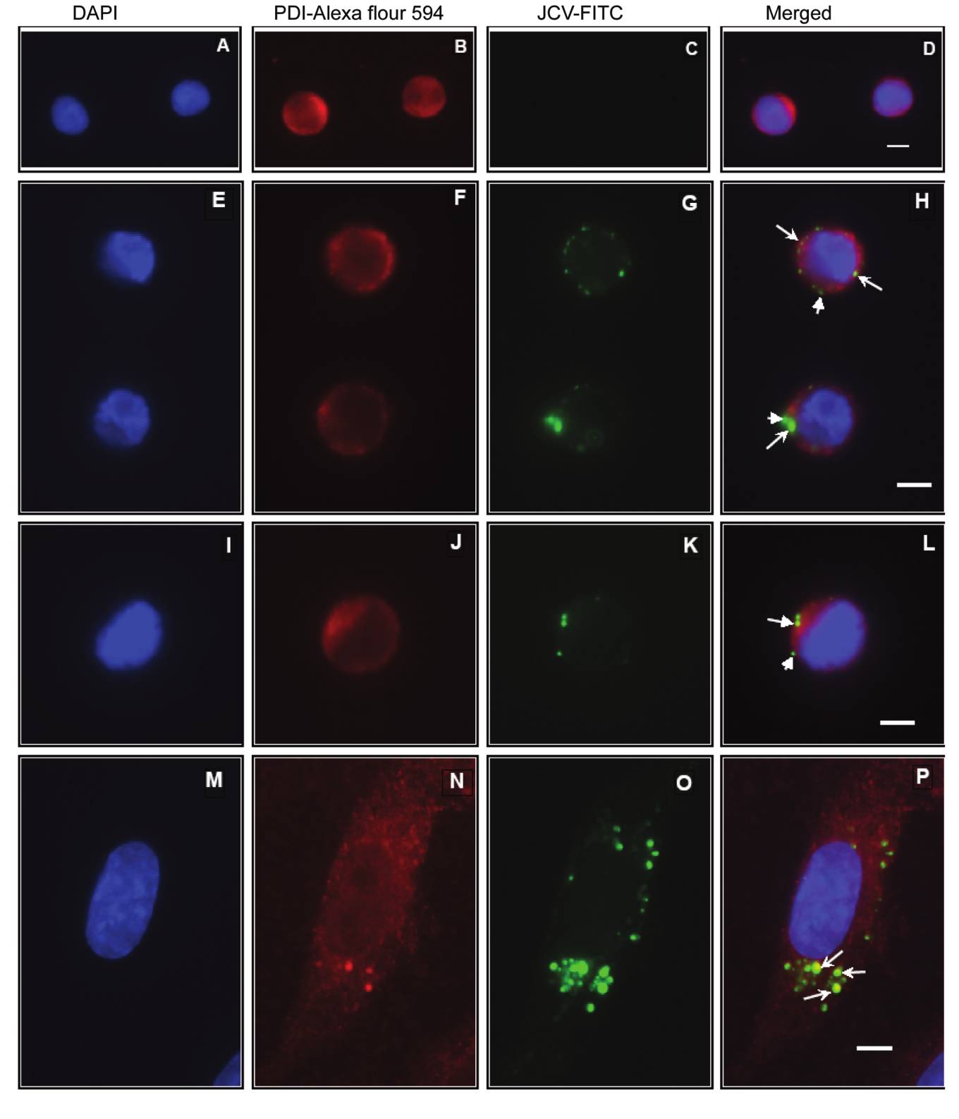
by 2 washes of PBS. Cells were cultured for up to 10 days. On We further characterized JCV infection of EBV-transformed days 1, 5, and 10, 2.5 ⫻ 10 trypsin-treated and untreated B B cells with use of FITC-labeled JCV. B cells and human brain cells were harvested, and JCV VP-1 DNA was amplified and microvascular endothelial (HBMVE) cells were either infect- quantitated by quantitative PCR. The data demonstrate that ed with FITC-labeled JCV (Figure 3E–3P) or inoculated with trypsin treatment had no significant effect in further reducing FITC-conjugated BSA (FITC-BSA) containing the same amount the JCV genome copies (Figure 1B), suggesting that JCV indeed of fluorescence (Figure 3A–3D) and were examined by immu- entered into the B cells.
nofluorescence microscopy. Interestingly, all FITC-labeled JCV Moreover, by infecting EBV-transformed B cells with FITC- virions were seen inside the HBMVE cells (Figure 3O–3P), labeled JCV and by employing flow cytometry, we confirmed whereas FITC-labeled JCV virions appeared to be inside the B that JCV infects B cells in a dose-dependent manner. When B cells and on the B cells surfaces (Figure 3G–3H and 3K–3L).
186 • JID 2010:202 (15 July) • Chapagain and Nerurkar


Figure 2.
JC virus (JCV) infection of B cells is dose-dependent. One million Epstein-Barr virus–transformed B cells were incubated with 125 (A), 250 (B ), 500 (C ), or 1000 (D ) HAU of fluorescein isothiocyanate (FITC)-JCV or with equivalent fluorescence-containing bovine serum albumin (AD ),similarly labeled with FITC (gray-filled histogram) as a control for 2 h, washed with phosphate-buffered saline, and subjected to flow cytometry.
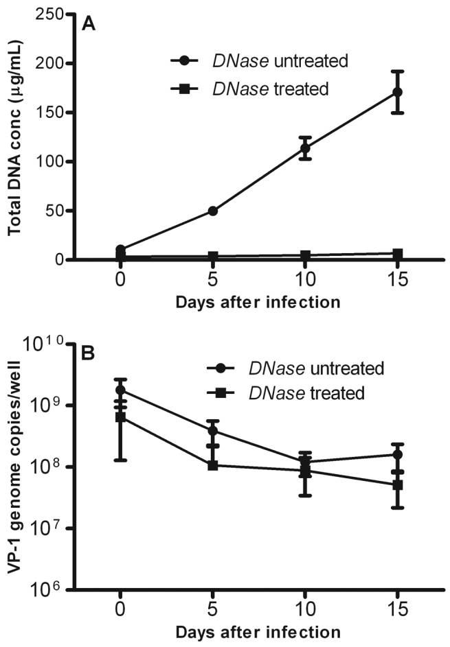
These data confirm that JCV indeed infects the B cells. However, DNase-treated and DNase-untreated cells, DNA concentration in contrast to HBMVE cells where VP-1 and T antigen proteins was measured, and JCV VP-1 DNA was quantitated by quan- were observed predominantly in the cell nucleus [32], our re- titative PCR. Figure 4A demonstrates that the total cellular DNA peated attempts to demonstrate T antigen and VP-1 proteins recovered from the B cells increased ∼16-fold (mean  stan- expression in the B cells failed, and it is unlikely that JCV infection dard deviation, 10.7  1.5 to 170.9  21.1 mg/mL) from days of B cells is productive. Collectively, our data demonstrate that 0 to 15 after infection, suggesting that the B cells were actively JCV infects the B cells; however, JCV infection of B cells is replicating. However, mean JCV VP-1 DNA copies/well ( standard deviation) decreased ∼11-fold ( 18.0  8.6 ⫻ 10 to Infectious JCV virions persist in the B cells.
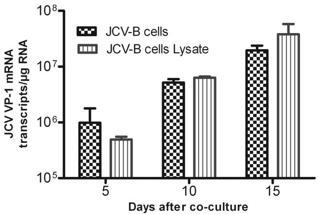
16.0  7.5 ⫻ 10 copies/well) from days 0 to 15 after infection, data suggest that JCV infects EBV-transformed B cells but does further confirming our observation that JCV infection of B not replicate in these cells, intact virus must survive inside the cells is nonproductive. Although DNase treatment was very B cells long enough to be trafficked across the blood-brain effective and reduced the total cellular DNA recovered from barrier and must then have a mechanism to be released to JCV-infected B cells, up to 96% (mean  standard deviation, infect susceptible cells, particularly the oligodendrocytes. How- 170.9  21.1 to 6.9  1.7 mg/mL) on day 15 after infection ever, our data suggest that JCV genome copy numbers rapidly (Figure 4A), DNase treatment had very little effect on the JCV decrease in the EBV-transformed B cells after infection, and it VP-1 DNA copies recovered from the B cells (Figure 4B). These was unclear whether the decrease in viral genome was the result results suggest that JCV virions in the B cells remain intact for of degradation of viral DNA inside the B cells or merely a result at least 15 days after infection and are protected from DNase of a dilution effect of replicating B cells, because in vitro EBV- transformed B cells replicate rapidly. To address this question, To further verify that JCV virions inside the B cells were 2.5 ⫻ 10 EBV-transformed B cells with 250 HAU infectious, JCV-infected B cells or lysates after infection of B of JCV for 2 h. After a wash, 2.5 ⫻ 10 cells were seeded in cells with JCV for 24 h were further cocultured for an additional each well of a 24-well plate, were cultured, and all cells from 24 h with naive PHFG cells. After 24 h coculture, the PHFG each well were harvested at each time point. The cells were cells were washed; cells were harvested at days 5, 10, and 15 freeze-thawed 4 times to mechanically lyse the cells, and trip- and were analyzed for viral late gene (VP-1) expression. Al- licate samples at each time point were either left untreated or though no viral transcripts were recovered from PHFG cells treated with 125 U/mL of DNase for 1 h at 37C to digest cocultured with uninfected B cells (data not shown), JCV VP- genomic and free viral DNA. The DNase-treated cells were heat 1 mRNA transcripts increased 1100-fold in PHFG cells cocul- inactivated at 75C for 10 min, DNA was extracted from both tured with JCV-infected B cells or JCV-infected B cells lysate, JCV Infection of Human B Lymphocytes • JID 2010:202 (15 July) • 187


Figure 3.
Epifluorescence microscopy demonstrating infection of B cells by fluorescein isothiocyanate (FITC)–conjugated JC virus (JCV). One million B cells were inoculated with either FITC-labeled bovine serum albumin (AD) as a negative control or 1000 HAU of FITC-labeled JCV (EL). Membraneswere labeled with protein disulphide isomerase (PDI) and visualized with Alexa flour 594 (red ). Cells were counterstained with 4,6-diamidino-2-phenylindole (DAPI) to visualize cell nuclei (blue). Merged images demonstrate FITC-labeled JCV on the B cell surface (arrowheads) and inside the Bcells (arrow ) (H and L). MP, Human brain microvascular endothelial cells infected with FITC-labeled JCV (positive control). Scale bar, 5 mm.


infected monocytes (Trojan horse) [42]. Further studies havealso suggested the presence of JCV DNA in the peripheral bloodmononuclear cells of 17.4% of 69 HIV type 1–infected im-munocompetent patients, in 23.2% of 82 HIV type 1–infectedimmunocompromised patients, and in 60% of AIDS patientswith PML [43]. However, there was no expression of JCV earlyand late mRNA transcripts in the peripheral blood mononu-clear cells [43]. Recently, JCV DNA was demonstrated in theperipheral blood mononuclear cells and VP-1–stained CD138+positive plasma cells were visualized in the bone marrow of apatient with rheumatoid arthritis treated with methotrexate,who developed PML and had a rapid fatal outcome, furthersuggesting that B lymphocytes may play an important role inthe JCV latency and dissemination [14, 16]. Similarly, JCV DNAwas detected in the bone marrow of HIV-negative and HIV–positive patients with and without PML, and JCV large T an-tigen but not VP1 was detected by double immunostaining inCD138+ plasma cells in an archival bone marrow specimenfrom an HIV-infected patient without PML [41]. Moreover,Focosi et al [44] detected JCV DNA in serial bone marrowsamples from 4 hematological patients with histology-con-firmed PML. Importantly, JCV DNA was first detected in thebone marrow and later in CSF, peripheral blood, and brain.
Furthermore, in 1 patient who survived, JCV was not detectedafter resolution of PML, suggesting that JCV indeed dissemi-nates by the hematogenous route [44].
It is often argued that JCV establishes low level of productive infection in B lymphocytes [8, 12, 18, 20]. Atwood et al [20] Figure 4.
JC virus (JCV) genome in B cells was virion protected. In- have demonstrated that 1% of B lymphocytes were JCV positive fected B cells harvested at different time points after infection were by in situ DNA hybridization on day 13 after infection, when mechanically lysed by repeated freeze-thaw and were untreated or treatedwith DNase. DNA was extracted, concentration was measured, and JCVVP-1 DNA was quantitated by quantitative polymerase chain reaction. A,Total cellular DNA recovered increased from day 0 to day 15, suggestingthat B cells were actively replicating and that DNase effectively reducedDNA concentration. B, JCV VP-1 DNA copies decreased from days 0 to15 after infection as expected, but DNase treatment had very little effecton the viral DNA, suggesting that JCV genome in the B cells was virionprotected from DNase digestion.
clearly indicating that the virions that remained inside the Bcell cytoplasm were infectious and replicated efficiently in naivePHFG cells (Figure 5).
The mechanism of JCV trafficking across the blood-brain bar-rier remains poorly understood. PCR analyses have demon- Figure 5.
JC virus (JCV)–infected B cells transmit JCV infection to strated that JCV may persist in the brain, tonsils, and lym- naive primary human fetal glial (PHFG) cells. PHFG cells were coculturedfor 24 h with JCV-infected B cells or B cells lysate, and JCV VP1 mes- phocytes of individuals with and without PML [2, 6–11, 13, senger RNA transcript expression in PHFG cells was quantitated. JCV 16, 40, 41], and it was proposed that JCV might employ B VP-1 messenger RNA transcripts increased several fold in PHFG cells lymphocytes to cross the blood-brain barrier [8, 12, 14–16], cocultured with JCV-infected B cells or B cells lysate. However, no viral similar to human immunodeficiency virus (HIV) and simian transcripts were detected from PHFG cells cocultured with uninfected B immunodeficiency virus, which gain entry into the brain via cells (data not shown).
JCV Infection of Human B Lymphocytes • JID 2010:202 (15 July) • 189
1 million B cells were infected with 10,000 HAU of JCV; they Although we always detected JCV TAg and VP-1 DNA, viral argued that it was indicative of DNA replication [20]. Monaco mRNA transcripts were never detected in JCV-infected B cells, et al [12] claimed that 3%–5% of hematopoietic cell line (KG- suggesting that JCV transcripts expression was either extremely 1a), primary hematopoietic progenitor cells (CD34+), and pri- low or completely shut off in the B cells. Further in vitro and mary B cells were positive for JCV on day 5 after infection in vivo studies using sensitive state-of-the-art real-time PCR when infected with 300 HAU/mL of JCV (Mad-4) [12]. Al- and quantitative reverse transcription PCR assays are warranted though TAg and VP-1 transcripts [18] and proteins [8] ex- to define the role of hematopetic precursor cells, primary B pression were demonstrated occasionally in JCV-infected B cell cells, and bone marrow–derived plasma cells in JCV replication, line or B lymphocytes in vitro, these studies were often limited latency, and dissemination.
to 1 time point after infection, or the transcripts were detectedonly after a second round of PCR (nested PCR) [18]. Taken together, in spite of several claims of replication of JCV genomeand production of infectious virions de novo in the B cells, We thank Dr Allison Imrie, University of Hawaii, for providing EBV- evidence for JCV replication in B lymphocytes is still lacking.
transformed primary B cells. We also thank Mr Nelson I. B. Lazaga, DrFrederic Mercier, Dr Pakieli Kaufusi, Ms Ulziijargal Gurjav, and Ms Al- In an attempt to better understand the mechanism(s) of JCV exandra Gurary for their technical assistance.
transmigration across the blood-brain barrier, we previouslydemonstrated that JCV productively infects HBMVE cells, prin-cipal cells lining the blood-brain barrier, and proposed that cell-free JCV may cross the blood-brain barrier by infecting 1. Astrom KE, Mancall EL, Richardson EP Jr. Progressive multifocal HBMVE cells [32]. Recently VP-1 expressing CD138+ plasma leuko-encephalopathy; a hitherto unrecognized complication of cells were demonstrated in the bone marrow with both rear- chronic lymphatic leukaemia and Hodgkin's disease. Brain 1958; 81:
93–111.
ranged and archetype regulatory regions, suggesting that B cells 2. Frisque RJ, White FA III. The molecular biology of JC virus, causative may latently harbor the virus and provide an environment for agent of progressive multifocal leukoencephalopathy. In: P. RR, ed.
emergence of rearranged form of JCV, and thus, may act as a Molecular neurovirology. Totowa, NJ: Humana Press, 1992:25–158.
3. Padgett BL, Walker DL, ZuRhein GM, Eckroade RJ, Dessel BH. Cul- vehicle for JCV dissemination [16]. However, the relative role tivation of papova-like virus from human brain with progressive mul- of B lymphocytes in JCV transmigration across the blood-brain tifocal leucoencephalopathy. Lancet 1971; 1:1257–1260.
barrier remains unclear. To address this issue, we employed 4. Richardson EP Jr. Our evolving understanding of progressive multifocal leukoencephalopathy. Ann N Y Acad Sci 1974; 230:358–364.
EBV-transformed B cells and studied the replication kinetics 5. Doerries K. Human Polyomavirus JC and BK persistent infection. In: of JCV. Although we did not find any evidence of JCV repli- Ahsan N, ed. Polyomaviruses and human diseases. Vol. 577: Springer cation in EBV-transformed B cells in vitro, we observed that Science and Landes Bioscience, 2006:102–116.
6. Dorries K, Vogel E, Gunther S, Czub S. Infection of human poly- JCV nonproductively infects EBV-transformed B cells and JCV omaviruses JC and BK in peripheral blood leukocytes from immu- virions remain intact inside the B cells, presumably long enough nocompetent individuals. Virology 1994; 198:59–70.
to employ B cells as a potential vehicle to cross the blood-brain 7. Major EO, Amemiya K, Tornatore CS, Houff SA, Berger JR. Patho- genesis and molecular biology of progressive multifocal leukoenceph- barrier and transmit infection to the oligodendrocytes in the alopathy, the JC virus-induced demyelinating disease of the human brain. Clin Microbiol Rev 1992; 5:49–73.
Our study does not preclude the possibility that primary B 8. Monaco MC, Jensen PN, Hou J, Durham LC, Major EO. Detection of JC virus DNA in human tonsil tissue: evidence for site of initial cells, CD34+ hematopetic precursor cells, or other cells within viral infection. J Virol 1998; 72:9918–9923.
this lineage would not support JCV replication. In fact, JCV 9. Monaco MC, Shin J, Major EO. JC virus infection in cells from lym- infection of primary hematopoietic CD34+ cells as well as he- phoid tissue. Dev Biol Stand 1998; 94:115–122.
matopoietic progenitor cell lines KG-1 and KG-1a has been 10. Tornatore C, Berger JR, Houff SA, et al. Detection of JC virus DNA in peripheral lymphocytes from patients with and without progressive suggested [9, 12]. However, there are no studies comparing multifocal leukoencephalopathy. Ann Neurol 1992; 31:454–462.
JCV replication between primary and EBV-transformed B cells; 11. White FA III, Ishaq M, Stoner GL, Frisque RJ. JC virus DNA is present interestingly, our data on EBV-transformed B cells are consis- in many human brain samples from patients without progressive mul-
tifocal leukoencephalopathy. J Virol 1992; 66:5726–5734.
tent with earlier studies that documented low efficiency of JCV 12. Monaco MC, Atwood WJ, Gravell M, Tornatore CS, Major EO. JC infection of primary B cells and hematopoietic precursor cells virus infection of hematopoietic progenitor cells, primary B lympho- [9, 12]. Moreover, JCV T antigen– and VP-1–expressing pri- cytes, and tonsillar stromal cells: implications for viral latency. J Virol
1996; 70:7004–7012.
mary B cells decreased from 1%–3% on day 1 to 0.5% on day 13. Newman JT, Frisque RJ. Detection of archetype and rearranged variants 8 after infection [9], suggesting that productive JCV replication of JC virus in multiple tissues from a pediatric PML patient. J Med is unlikely to occur even in primary B cells and hematopetic Virol 1997; 52:243–252.
14. Houff SA, Berger JR. The bone marrow, B cells, and JC virus. J Neu- precursor cells. These studies used less sensitive immunostain- rovirol 2008; 14:341–343.
ing, HA assay, or standard PCR to determine JCV infection.
15. Houff SA, Major EO, Katz DA, et al. Involvement of JC virus-infected 190 • JID 2010:202 (15 July) • Chapagain and Nerurkar
mononuclear cells from the bone marrow and spleen in the patho- 31. Chapagain ML, Nguyen T, Bui T, Verma S, Nerurkar VR. Comparison genesis of progressive multifocal leukoencephalopathy. N Engl J Med of real-time PCR and hemagglutination assay for quantitation of hu- man polyomavirus JC. Virol J 2006; 3:3.
16. Marzocchetti A, Wuthrich C, Tan CS, et al. Rearrangement of the JC 32. Chapagain ML, Verma S, Mercier F, Yanagihara R, Nerurkar VR. Poly- virus regulatory region sequence in the bone marrow of a patient with omavirus JC infects human brain microvascular endothelial cells inde- rheumatoid arthritis and progressive multifocal leukoencephalopathy.
pendent of serotonin receptor 2A. Virology 2007; 364:55–63.
J Neurovirol 2008; 14:455–458.
33. Co JK, Verma S, Gurjav U, Sumibcay L, Nerurkar VR. Interferon- alpha 17. Sabath BF, Major EO. Traffic of JC virus from sites of initial infection and- beta restrict polyomavirus JC replication in primary human fetal to the brain: the path to progressive multifocal leukoencephalopathy.
glial cells: implications for progressive multifocal leukoencephalopathy J Infect Dis 2002; 186(suppl 2):S180–S186.
therapy. J Infect Dis 2007; 196:712–718.
18. Wei G, Liu CK, Atwood WJ. JC virus binds to primary human glial 34. Verma S, Ziegler K, Ananthula P, et al. JC virus induces altered patterns cells, tonsillar stromal cells, and B-lymphocytes, but not to T lym- of cellular gene expression: Interferon-inducible genes as major tran- phocytes. J Neurovirol 2000; 6:127–136.
scriptional targets. Virology 2006; 345:457–467.
19. Major EO, Amemiya K, Elder G, Houff SA. Glial cells of the human 35. Alter A, Duddy M, Hebert S, et al. Determinants of human B cell developing brain and B cells of the immune system share a common migration across brain endothelial cells. J Immunol 2003; 170:4497–
DNA binding factor for recognition of the regulatory sequences of the human polyomavirus, JCV. J Neurosci Res 1990; 27:461–471.
36. Imrie A, Meeks J, Gurary A, et al. Differential functional avidity of 20. Atwood WJ, Amemiya K, Traub R, Harms J, Major EO. Interaction dengue virus-specific T-cell clones for variant peptides representing of the human polyomavirus, JCV, with human B-lymphocytes. Virology heterologous and previously encountered serotypes. J Virol 2007; 81:
21. Kleinschmidt-DeMasters BK, Tyler KL. Progressive multifocal leuko- 37. Liu CK, Hope AP, Atwood WJ. The human polyomavirus, JCV, does encephalopathy complicating treatment with natalizumab and inter- not share receptor specificity with SV40 on human glial cells. J Neu- feron beta-1a for multiple sclerosis. N Engl J Med 2005; 353:369–374.
rovirol 1998; 4:49–58.
22. Langer-Gould A, Atlas SW, Green AJ, Bollen AW, Pelletier D. Pro- 38. Liu CK, Wei G, Atwood WJ. Infection of glial cells by the human gressive multifocal leukoencephalopathy in a patient treated with na- polyomavirus JC is mediated by an N-linked glycoprotein containing talizumab. N Engl J Med 2005; 353:375–381.
terminal alpha(2–6)-linked sialic acids. J Virol 1998; 72:4643–4649.
23. Van Assche G, Van Ranst M, Sciot R, et al. Progressive multifocal 39. Chapagain ML, Sumibcay L, Gurjav U, Kaufusi PH, Kast RE, Nerurkar leukoencephalopathy after natalizumab therapy for Crohn's disease. N VR. Serotonin receptor 2A blocker (risperidone) has no effect on hu- Engl J Med 2005; 353:362–368.
man polyomavirus JC infection of primary human fetal glial cells. J 24. Rice GP, Hartung HP, Calabresi PA. Anti-alpha4 integrin therapy for Neurovirol 2008; 14:448–454.
multiple sclerosis: mechanisms and rationale. Neurology 2005; 64:1336–
40. Newman JT, Frisque RJ. Identification of JC virus variants in multiple tissues of pediatric and adult PML patients. J Med Virol 1999; 58:79–86.
25. Arroyo AG, Yang JT, Rayburn H, Hynes RO. Differential requirements for alpha4 integrins during fetal and adult hematopoiesis. Cell 1996;
41. Tan CS, Dezube BJ, Bhargava P, et al. Detection of JC virus DNA and proteins in the bone marrow of HIV-positive and HIV-negative pa- 26. Ransohoff RM. Natalizumab and PML. Nat Neurosci 2005; 8:1275.
tients: implications for viral latency and neurotropic transformation.
27. Ransohoff RM. "Thinking without thinking" about natalizumab and J Infect Dis 2009; 199:881–888.
PML. J Neurol Sci 2007; 259:50–52.
42. Georgsson G. Neuropathologic aspects of lentiviral infections. Ann N 28. Genetech. Important drug warning regarding Raptiva (efalizumab).
Y Acad Sci 1994; 724:50–67.
43. Andreoletti L, Dubois V, Lescieux A, et al. Human polyomavirus JC raptiva_dhcp_0309.pdf. Accessed 10 April 2009.
latency and reactivation status in blood of HIV-1-positive immuno- 29. Krueger JG. The immunologic basis for the treatment of psoriasis with compromised patients with and without progressive multifocal leu- new biologic agents. J Am Acad Dermatol 2002; 46:1–23; quiz, 23–26.
koencephalopathy. AIDS 1999; 13:1469–1475.
30. Lebwohl M, Tyring SK, Hamilton TK, et al. A novel targeted T-cell 44. Focosi D, Maggi F, Andreoli E, Lanini L, Ceccherini-Nelli L, Petrini modulator, efalizumab, for plaque psoriasis. N Engl J Med 2003; 349:
M. The role of bone marrow cells for JCV pathogenicity. J Clin Virol JCV Infection of Human B Lymphocytes • JID 2010:202 (15 July) • 191

Source: http://files.immunologyresearch.ch/ial/ial-aricle-chapagain-jid-2010.pdf

sbts.edu

& the Authority of Christ by Russell D. Moore Counseling and the Authority of Christ: A New Vision for Biblical Counseling at The Southern Baptist Theological Seminary "And they were astonished at His teaching, for He taught them as one having authority, and not as the scribes." [Mark 1:22 ESV] The story of The Southern Baptist Theological Seminary is seen most clearly not in

Power from the sun 11-2014_gb.indd

Power from the sun Free solar energy for generating power using photovoltaics As renewable energy systems have become more widespread, consumers and end users have become increasingly keen to generate their own electricity. Today, an efficient photovoltaic system offers the opportunity to make profitable use of free solar energy. By installing photovoltaic modules, users demonstrate their