Levitra enthält Vardenafil, das eine kürzere Wirkdauer als Tadalafil hat, dafür aber schnell einsetzt. Männer, die diskret bestellen möchten, suchen häufig nach levitra kaufen ohne rezept. Dabei spielt die rechtliche Lage in der Schweiz eine wichtige Rolle.
06_077_088_phyex8_ap_ch06
06_077_088_PhyEx8_AP_Ch06 1/10/08 5:23 PM Page 77
Frog Cardiovascular Physiology
O B J E C T I V E S
1. To list the properties of cardiac muscle as automaticity and rhythmicity,
and to define each.
2. To explain the statement, "Cardiac muscle has an intrinsic ability to
3. To compare the relative length of the refractory period of cardiac muscle
with that of skeletal muscle, and to explain why it is not possible totetanize cardiac muscle.
4. To define
extrasystole, and to explain at what point in the cardiac cycle
(and on an ECG tracing) an extrasystole can be induced.
5. To describe the effect of the following on heart rate: vagal stimulation,
cold, heat, pilocarpine, atropine, epinephrine, digitalis, and potassium,sodium, and calcium ions.
6. To define
vagal escape and discuss its value.
7. To define
ectopic pacemaker.
Investigation of human cardiovascular physiology is very interesting, but many
areas obviously do not lend themselves to experimentation. It would be tanta-mount to murder to inject a human subject with various drugs to observe their
effects on heart activity or to expose the human heart in order to study the lengthof its refractory period. However, this type of investigation can be done on frogsor computer simulations and provides valuable data because the physiologicalmechanisms in these animals, or programmed into the computer simulation, aresimilar if not identical to those in humans.
In this exercise, you will conduct the cardiac investigations just mentioned.
Special Electrical Properties
of Cardiac Muscle: Automaticity
and Rhythmicity
Cardiac muscle differs from skeletal muscle both functionally and in its fine struc-
ture. Skeletal muscle must be electrically stimulated to contract. In contrast, heart
muscle can and does depolarize spontaneously in the absence of external stimula-
tion. This property, called
automaticity, is due to plasma membranes that have re-
duced permeability to potassium ions but still allow sodium ions to slowly leak
into the cells. This leakage causes the muscle cells to slowly depolarize until the
action potential threshold is reached and
fast calcium channels open, allowing
Ca2+ entry from the extracellular fluid. Shortly thereafter, contraction occurs.
The spontaneous depolarization-repolarization events occur in a regular and
continuous manner in cardiac muscle, a property referred to as
rhythmicity.
In the following experiment, you will observe these properties of cardiac
muscle in a computer simulation. Additionally, your instructor may demonstratethis procedure using a real frog.
06_077_088_PhyEx8_AP_Ch06 1/10/08 5:23 PM Page 78
Nervous Stimulation
pulses fail to reach the ventricles (as in heart block), the ven-tricles continue to beat but at their own inherent rate, which is
of the Heart
much slower than that usually imposed on them. Althoughheart contraction does not depend on nerve impulses, its rate
Both the parasympathetic and sympathetic nervous systems in-
can be modified by extrinsic impulses reaching it through the
nervate the heart. Stimulation of the sympathetic nervous sys-
autonomic nerves. Cardiac activity is also modified by vari-
tem increases the rate and force of contraction of the heart. Stim-
ous chemicals, hormones, ions, and metabolites. The effects
ulation of the parasympathetic nervous system (vagal nerves)
of several of these chemical factors are examined in the next
decreases the depolarization rhythm of the sinoatrial node and
experimental series, Activities 4–9.
slows transmission of excitation through the atrio-ventricular
The frog heart has two atria and a single, incompletely
node. If vagal stimulation is excessive, the heart will stop beat-
divided ventricle. The pacemaker is located in the sinus veno-
ing. After a short time, the ventricles will begin to beat again.
sus, an enlarged region between the venae cavae and the right
This is referred to as vagal escape and may be the result of sym-
atrium. The sinoatrial (SA) node of mammals may have
pathetic reflexes or initiation of a rhythm by the Purkinje fibers.
evolved from the sinus venosus.
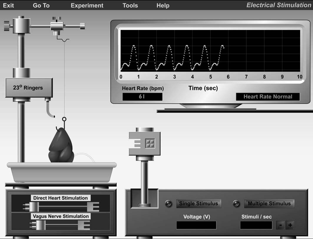
Choose Exercise 6: Frog Cardiovascular Physiology
Baseline Frog Heart Activity
from the drop-down menu and click GO. Then click Electri-
cal Stimulation. The opening screen will appear in a few
seconds (Figure 6.1). When the program starts, you will see a
The heart's effectiveness as a pump is dependent both on in-
tracing of the frog's heartbeat on the oscilloscope display in
trinsic (within the heart) and extrinsic (external to the heart)
the upper right part of the screen. Because the simulation au-
controls. In the first experimental series, Activities 1–3, you
tomatically adjusts itself to your computer's speed, you may
will investigate some of these factors.
not see the heart tracing appear in real time. If you want to
The nodal system, in which the "pacemaker" imposes its
increase the speed of the tracing (at the expense of tracing
depolarization rate on the rest of the heart, is one intrinsic
quality), click the Tools menu, choose Modify Display, and
factor that influences the heart's pumping action. If its im-
then select Increase Speed.
F I G U R E 6 . 1 Opening screen of the Electrical Stimulation experiment.
06_077_088_PhyEx8_AP_Ch06 1/10/08 5:23 PM Page 79
Frog Cardiovascular Physiology
To familiarize yourself with the equipment, choose
Record the number of ventricular contractions per minute
Balloons On/Off from the Help menu. This feature allows
displayed in the Heart Rate window under the oscilloscope.
you to scroll around the screen and view equipment labels.
You can turn off this feature by returning to the Help menu
bpm (beats per minute) ■
and selecting Balloons On/Off.
The oscilloscope display shows the ventricular contraction
A C T I V I T Y
rate in the Heart Rate window. The heart activity window to theright of the Heart Rate display provides the following messages:
Investigating the Refractory Period
• Heart Rate Normal—displayed when the heart is beating
of Cardiac Muscle
under resting conditions.
In Exercise 2 you saw that repeated rapid stimuli could cause
• Heart Rate Changing—displayed when the heart rate is
skeletal muscle to remain in a contracted state. In other
increasing or decreasing.
words, the muscle could be tetanized. This was possible be-
• Heart Rate Stable—displayed when the heart rate is
cause of the relatively short refractory period of skeletal mus-
steady, but higher or lower than normal. For example, if you
cle. In this experiment you will investigate the refractory pe-
applied a chemical that increased heart rate to a stable but
riod of cardiac muscle and its response to stimulation.
higher-than-normal rate, you would see this message.
Click and hold the mouse button on the Direct Heart
Stimulation electrode, and drag it to the electrode holder.
The electrical stimulator is below the oscilloscope display. In
the experiment, clicking Single Stimulus delivers a single
Release the mouse button to lock the electrode in place.
electrical shock to the frog heart. Clicking Multiple Stimulus
The electrode will touch the ventricular muscle tissue.
delivers repeated electrical shocks at the rate indicated in the
Deliver single shocks in succession by clicking Single
Stimuli/sec window just below the Multiple Stimulus button.
Stimulus rapidly. You may need to practice to acquire the
When the Multiple Stimulus button is clicked, it changes to a
correct technique.
Stop Stimulus button that allows you to stop electrical stimu-
lation as desired. Clicking the (+) or (⫺) buttons next to the
Watch for extrasystoles, which are extra beats that show
Stimuli/sec window adjusts the stimulus rate. The voltage de-
up riding on the ventricular contraction peak. Also note the
livered when Single Stimulus or Multiple Stimulus is clicked
compensatory pause, which allows the heart to get back on
is displayed in the Voltage window just below the Single
schedule after an extrasystole (Figure 6.2b).
Stimulus button. The simulation automatically adjusts thevoltage for the experiment. The postlike apparatus extendingupward from the electrical stimulator is the electrode holderinto which you will drag-and-drop electrodes from the supply
Ventricular systole
cabinet in the bottom left corner of the screen.
The left side of the screen contains the apparatus that
Ventricular diastole
sustains the frog heart. The heart has been lifted away fromthe body of the frog by a hook passed through the apex of theheart. Although the frog cannot be seen because it is in thedissection tray, its heart has not been removed from its circu-latory system. A thin string connects the hook in the heart tothe force transducer at the top of the support bracket. As theheart contracts, the string exerts tension on the force trans-
One-second time line
ducer, which converts the contraction into the oscilloscopetracing. The slender white strand extending from the heart to-
ward the right side of the dissection tray is the vagus nerve. In
the simulation, room-temperature (23°C) frog Ringer's solu-tion continuously drips onto the heart to keep it moist and re-
sponsive so that a regular heart beat is maintained.
The two electrodes you will use during the experiment are
located in the supply cabinet beneath the dissection tray. The
Direct Heart Stimulation electrode is used to stimulate the
ventricular muscle directly. The Vagus Nerve Stimulation
electrode is used to stimulate the vagus nerve. To position ei-
One-second time line
ther electrode, click and drag the electrode to the two-prongedplug in the electrode holder and then release the mouse button.
F I G U R E 6 . 2 Recording of contractile activity of
a frog heart. (a) Normal heartbeat. (b) Induction of an
A C T I V I T Y
Recording Baseline Frog Heart Activity
Before beginning to stimulate the frog heart experimen-
tally, watch several heartbeats. Be sure you can distinguishatrial and ventricular contraction (Figure 6.2a).
06_077_088_PhyEx8_AP_Ch06 1/10/08 5:23 PM Page 80
On the basis of the recording, during which portion of the
Assessing Physical and
cardiac cycle was it possible to induce an extrasystole? UseFigures 6.2a and b to help you decide.
Chemical Modifiers
of Heart Rate
Now that you have observed normal frog heart activity, you
Attempt to tetanize the heart by clicking Multiple Stim-
will have an opportunity to investigate the effects of various
ulus. Electrical shocks will be delivered to the muscle at a
modifying factors on heart activity. After removing the agent
rate of 20 stimuli/sec. What is the result?
in each activity, allow the heart to return to its normal rate be-fore continuing with the testing.
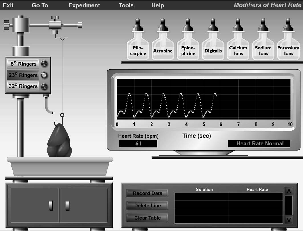
Choose Modifiers of Heart Rate from the Experiment
menu. The opening screen will appear in a few seconds(Figure 6.3). The appearance and functionality of the os-cilloscope display is the same as it was in the Electrical Stim-
Considering the function of the heart, why is it important that
ulation experiment. The solutions shelf above the oscillo-
the heart muscle cannot be tetanized?
scope display contains the chemicals you'll use to modifyheart rate in the experiment. You can choose the temperatureof the Ringer's solution dispensed by clicking the appropriatebutton in the Ringer's dispenser at the left part of the screen.
The doors to the supply cabinet are closed during this exper-iment because the electrical stimulator is not used.
When you click Record Data in the data control unit
Click Stop Stimulus to stop the electrical stimulation. ■
below the oscilloscope, your data is stored in the computer'smemory and is displayed in the data grid at the bottom of the
A C T I V I T Y
screen; data displayed include the solution used and the re-sulting heart rate. If you are not satisfied with a trial, you can
Examining the Effect
click Delete Line. Click Clear Table if you wish to repeat
of Vagus Nerve Stimulation
the entire experiment.
The vagus nerve carries parasympathetic impulses to theheart, which modify heart activity.
A C T I V I T Y
Click the Direct Heart Stimulation electrode to return
Assessing the Effect of Temperature
it to the supply cabinet.
Predict what effect a decrease in temperature will have on
Click and drag the Vagus Nerve Stimulation electrode
heart rate and write your prediction below.
to the electrode holder.
Release the mouse button to lock the electrode in place.
The vagus nerve will automatically be draped over the elec-trode contacts.
Click the 5°C Ringer's button to bathe the frog heart in
cold Ringer's solution. Watch the recording for a change in
Adjust the stimulator to 50 stimuli/sec by clicking the
cardiac activity.
(+) or (⫺) buttons.
When the heart activity window displays the message
Click Multiple Stimulus. Allow the vagal stimulation
Heart Rate Stable, click Record Data to retain your data in
to continue until the heart stops momentarily and then
the data grid.
begins to beat again (vagal escape), and then click Stop
Stimulus.
What change occurred with the cold (5°C) Ringer's solution?Compare to the baseline value recorded in Activity 1.
What is the effect of vagal stimulation on heart rate?
Did this change match your prediction? _
Now click the 23°C Ringer's button to flood the heart
with fresh room-temperature Ringer's solution.
The phenomenon of vagal escape demonstrates that many
After you see the message Heart Rate Normal in the
factors are involved in heart regulation and that any deleteri-
heart activity window, click the 32°C Ringer's button.
ous factor (in this case, excessive vagal stimulation) will be
When the heart activity window displays the message
overcome, if possible, by other physiological mechanisms
Heart Rate Stable, click Record Data to retain your data.
such as activation of the sympathetic division of the auto-nomic nervous system (ANS). ■
06_077_088_PhyEx8_AP_Ch06 1/10/08 5:23 PM Page 81
Frog Cardiovascular Physiology
F I G U R E 6 . 3 Opening screen of the Modifiers of Heart Rate experiment.
What change occurred with the warm (32⬚C) Ringer's
A C T I V I T Y
Assessing the Effect of Pilocarpine
Click and hold the mouse on the pilocarpine dropper cap.
Drag the dropper cap to a point about an inch above the
Record the heart rate at the two temperatures below.
heart, and release the mouse.
bpm at 5⬚C; bpm at 32⬚C
Pilocarpine solution will be dispensed onto the heart,
and the dropper cap will automatically return to the pilo-carpine bottle.
What can you say about the effect of temperature on heart rate?
Watch the heart activity window for the message Heart
Rate Stable, indicating that the heart rate has stabilized underthe effects of pilocarpine.
After the heart rate stabilizes, record the heart rate in the
space provided below, and click Record Data to retain your
Click the 23°C Ringer's button to flush the heart with
data in the grid.
fresh Ringer's solution. Watch the heart activity windowfor the message Heart Rate Normal before beginning the
06_077_088_PhyEx8_AP_Ch06 1/10/08 5:23 PM Page 82
What happened when the heart was bathed in the pilocarpine
Watch the heart activity window for the message Heart
Rate Stable.
After the heart rate stabilizes, record the heart rate in the
space provided below, and click Record Data to retain your
data in the grid.
Click the 23°C Ringer's button to flush the heart with
fresh Ringer's solution. Watch the heart activity window for
the message Heart Rate Normal, an indication that the heartis ready for the next test. ■
What happened when the heart was bathed in the epinephrine
Pilocarpine simulates the effect of parasympathetic nerve
(hence, vagal) stimulation by enhancing acetylcholine re-lease; such drugs are called parasympathomimetic drugs.
A C T I V I T Y
Which division of the autonomic nervous system does its ef-fect imitate?
Assessing the Effect of Atropine
Drag-and-drop the atropine dropper cap to a point about
an inch above the heart.
Click the 23°C Ringer's button to flush the heart with
Atropine solution will automatically drip onto the heart,
fresh Ringer's solution. Watch the heart activity window for
and the dropper cap will return to its position in the atropine
the message Heart Rate Normal, meaning that the heart is
ready for the next test. ■
Watch the heart activity window for the message Heart
Rate Stable.
A C T I V I T Y
After the heart rate stabilizes, record the heart rate in the
space below, and click Record Data to retain your data in the
Assessing the Effect of Digitalis
Drag-and-drop the digitalis dropper cap to a point about
an inch above the heart.
Digitalis solution will automatically drip onto the heart,
What is the effect of atropine on the heart?
and then the dropper will return to the digitalis bottle.
Watch the heart activity window to the right of the Heart
Rate window for the message Heart Rate Stable.
After the heart rate stabilizes, record the heart rate in the
Atropine is a drug that blocks the effect of the neurotransmit-
space provided below, and click Record Data to retain your
ter acetylcholine, liberated by the parasympathetic nerve
data in the grid.
endings. Do your results accurately reflect this effect of at-ropine?
What is the effect of digitalis on the heart?
Are pilocarpine and atropine agonists or antagonists with re-spect to each other in their effects on heart activity?
Click the 23°C Ringer's button to flush the heart with
fresh Ringer's solution. Watch the heart activity window for themessage Heart Rate Normal, then proceed to the next test. ■
Click the 23°C Ringer's button to flush the heart with
fresh Ringer's solution. Watch the heart activity window for
Digitalis (also known as digoxin and digitoxin) is a drug
the message Heart Rate Normal before beginning the next
commonly prescribed for heart patients with congestive heart
failure. It slows heart rate and strengthens the force of con-traction of the heart, providing more time for venous return
A C T I V I T Y
and decreasing the workload on the weakened heart. Theseeffects are thought to be due to inhibition of the sodium-
Assessing the Effect of Epinephrine
potassium pump and enhancement of Ca2+ entry into myo-cardial fibers.
Drag-and-drop the epinephrine dropper cap to a point
about an inch above the heart.
Epinephrine solution will be dispensed onto the heart,
and the dropper cap will return to the epinephrine bottle.
06_077_088_PhyEx8_AP_Ch06 1/10/08 5:23 PM Page 83
Frog Cardiovascular Physiology
A C T I V I T Y
Effect of K⫹:
Assessing the Effect of Various Ions
Describe what happened to the recording.
To test the effect of various ions on the heart, apply the de-sired solution using the following method.
Drag-and-drop the calcium ions dropper cap to a point
about an inch above the heart.
Describe your observations of the rhythm of the heartbeat.
Calcium ions will automatically be dripped onto the heart,
and the dropper cap will return to the calcium ions bottle.
Watch the heart activity window for the message Heart
Rate Stable.
When you see Heart Rate Stable on the screen, record
the heart rate in the space provided below step 6, and click
Potassium ion concentration is normally higher within cells
Record Data to retain your data in the grid.
than in the extracellular fluid. Hyperkalemia decreases the
Click the 23°C Ringer's button to flush the heart with
resting potential of plasma membranes, thus decreasing the
fresh Ringer's solution. Watch the heart activity window for
force of heart contraction. In some cases, the conduction rate
the message Heart Rate Normal, which means that the heart
of the heart is so depressed that ectopic pacemakers
is ready for the next test.
(pacemakers appearing erratically and at abnormal sites inthe heart muscle) appear in the ventricle, and fibrillation
Repeat steps 1 through 5 for sodium ions and then
Was there any evidence of premature beats in the recording of
potassium ion effects?
Does the heart rate stabilize and remain stable?
Was arrhythmia produced with any of the ions tested?
Describe your observations of the rhythm of the heartbeat.
Click Tools → Print Data to print your recorded data for
this experiment. ■
Effect of Na⫹:
Histology Review Supplement
Does the heart rate stabilize and remain stable?
For a review of cardiovascular tissue, go to Exercise H:
Histology Atlas and Review on the PhysioEx website to
print out the Cardiovascular Tissue Review worksheet.
Describe your observations of the rhythm of the heartbeat.
Source: http://www.clover.k12.sc.us/site/handlers/filedownload.ashx?moduleinstanceid=13857&dataid=9215&FileName=worksheet_ap06.pdf
INTEGRATED PEST MANAGEMENT PACKAGE M Srinivas Prasad National Centre for Integrated Pest Management LBS Building, IARI Campus, New Delhi – 110 012 Directorate of Plant Protection, National Institute of Plant Health Quarantine & Storage (DPPQ&S) CGO Complex, NH IV, Faridabad DAC, Min of Agri., Rajendranagar, Hyderabad- 500030
January - March 2010 / # 1 2 hours ago our wandering female harrier from the west In this issue: coast flew straight into the heart of Lesotho! She is now close to a smal stream in a high altitude val ey, about 1 More travels by Lockie . . . . . . Rob Simmons 32 km from the Katse Dam. She has flown a straight line distance of 1173 km from her coastal "home" near