Metformin mitigates the impaired development of skeletal muscle in the offspring of obese mice
Citation: Nutrition and Diabetes (2011) 1, e7; doi:10.1038/nutd.2011.3& 2011 Macmillan Publishers Limited All rights reserved 2044-4052/11
Metformin mitigates the impaired development ofskeletal muscle in the offspring of obese mice
JF Tong1, X Yan1, JX Zhao1, MJ Zhu1, PW Nathanielsz2 and M Du1
1Developmental Biology Group, Department of Animal Science, University of Wyoming, Laramie, WY, USA and 2Center forPregnancy and Newborn Research, University of Texas Health Sciences Center, San Antonio, TX, USA
Background: Maternal obesity is linked with offspring obesity and type 2 diabetes. Skeletal muscle (SM) insulin resistance iscentral to the development of diabetes. Adenosine monophosphate (AMP)-activated protein kinase (AMPK) is inhibited in SM offetuses born to obese mothers.
Objective: The aim of this study was to evaluate the effect of maternal metformin administration on AMPK activity and reversionof adverse changes in offspring SM of obese mice.
Design: Female weanling C57BL/6J mice received either control diet (CON, 6 mice) or high-fat diet (HFD; OB, 12 mice) for 8weeks before mating. After mating, mice continued receiving their respective CON or OB diets. In addition, 6 of those 12 micefed with fat diet also received metformin administration (2 mg per ml in drinking water) throughout gestation and lactation(MET). After weaning at postnatal 21 days, offspring were fed a HFD to mimic a postnatal obesogenic environment untilnecropsy.
Results: Mothers receiving the fat diet developed obesity. OB offspring showed higher adiposity than CON and MET offspring.
AMPK phosphorylation was lower in SM of OB offspring. b-Catenin and myogenic regulatory factors, MyoD and myogenin, weredownregulated in OB muscle, whereas the adipogenic marker, peroxisome proliferator-activated receptor-g, was upregulatedcompared with CON muscle. Metformin administration prevented these changes in OB offspring SM. OB but not MET offspringdemonstrated glucose intolerance. Mitochondrial content decreased, and activities of citrate synthase and b-hydroxyacyl-CoAdehydrogenase also decreased in OB offspring SM, whereas they were recovered in MET offspring SM.
Conclusion: Maternal metformin administration improves SM development in OB offspring.
Nutrition and Diabetes (2011) 1, e7; published online 16 May 2011
Keywords: developmental programming; maternal obesity; Amp-activated protein kinase; metformin; skeletal muscle
Skeletal muscle (SM) is a potential target for antiobese and
antidiabetic therapies because it constitutes about 40–50% of
Obesity and type 2 diabetes are increasing at alarming rates,
body mass and is the main peripheral tissue responsive to
even in teenagers and At the same time, the
insulin-stimulated uptake of glucose and fatty acids.
prevalence of overweight and obese women of childbearing
Development of insulin resistance in SM is an essential step
age is a growing public health concern.Western high-
in the development of type 2 The fetal stage is
energy diets combined with maternal obesity (MO) represent
crucial for SM development, because no net increase in the
a special problem that can result in harmful, persistent
number of muscle fibers occurs after
phenotypic outcomes in offspring, including predisposition
Fetal SM contains a large number of pluripotent cells.
to obesity and Although accumulating evidence
Although the majority of these cells differentiate into muscle
from both epidemiological studies and animal studies shows
fibers through myogenesis, a portion becomes adipocytes.
that MO predisposes offspring to obesity and diabetes,
The canonical Wnt/b-catenin signaling pathway is essential
the causative mechanisms have not been elucidated.
for myogenesis.Enhancing Wnt/b-catenin signalingincreases expression of myogenic regulatory factors, includ-ing Myf5 and MyoD,whereas downregulating Wnt/
Correspondence: Dr M Du, Developmental Biology Group, Department of
b-catenin signaling pathway promotes adipogenesis by
Animal Science, University of Wyoming, 1000 East University Avenue,
enhancing the expression of peroxisome proliferator-
Laramie, WY 82071, USA.
activated receptor-g (PPAR-g), a key regulator of adipogenesi
E-mail: Received 4 August 2010; revised 25 March 2011; accepted 27 March 2011
Enhanced adipogenesis in fetal SM increases intramuscular
Maternal obesity, metformin and offspring muscle
adipocytes, an event linked to SM insulin resisThus,
At weaning, maternal mice were killed, and serum was
impairment of fetal SM development will predispose offspring
collected for blood profile analyses. The maternal carcasses
to diabetes and obesity in later
were grounded, and the fat content was analyzed using Folch
Adenosine monophosphate (AMP)-activated protein kinase
extraction method.After weaning, all offspring were fed
(AMPK) is a master controller of energy metabolism in
with the HFD (D12451) to mimic a postnatal obesogenic
SM.AMPK controls SM development by enhancing the
environment until necropsy at 60 days of age.
A separate cohort of 12 female mice were assigned to CON,
genesis,both of which link to SM insulin resistance.
OB and MET groups (n ¼ 4) and treated as described above.
Activation of AMPK promotes myogenesis, whereas inhibition
Offspring male mice were used for glucose tolerance test.
promotes adipogenesisMetformin is the most commondrug currently in use for treating type 2 diabetes. Metformin'sactivity is mainly mediated by AMPKUsing a maternal
Sample collection
obese sheep model, we have showed that MO downregulates
Offspring were killed on postnatal day 1 (two mice per litter
AMPK activity in fetal SM, which is expected to have
were killed), day 15 (one male per litter) and day 60 (one
profound impact on glucose uptake, insulin signaling and
male per litter). Each mouse was skinned and eviscerated.
properties of fetal and offspring SM.Based on these findings,
Owing to the small size of the offspring, the muscle from the
we hypothesized that activation of fetal SM AMPK by
whole hind (after removing skin, bone and visible connec-
maternal metformin administration would prevent the
tive tissue and fat) was collected from postnatal day-1 mice
changes in fetal and offspring SM properties that result from
(muscles from 2 mice per litter were pooled) and day-15 male
exposure to MO. The objective of the present study was to
mice (1 mouse per litter, n ¼ 6 per treatment), frozen in
investigate the effects of metformin administration to obese
liquid N2 and stored at 80 1C for further analyses. For
mothers on offspring SM composition and to assess the
postnatal day-60 mice, gastrocnemius muscle and gonadal
importance of AMPK in the development of SM.
fat from males (n ¼ 6 per group) were separated, weighed andcollected. Also, a section (from first to last lumbar vertebrae)
Materials and methods
of trunk from day-1 and -15 mice was embedded in OCTcompound (Sakura Fineteck USA Inc., Torrance, CA, USA),
Care and use of animals
frozen in liquid N2 and stored in 80 1C for histological
examination of muscle structure.
Wilmington, MA, USA) were maintained according to thestandard protocols approved by the University of Wyoming
Animal Use and Care Committee. Mice were housed two per
Antibodies against AMPKa (cat. number 2532), phospho-
cage under a 12-h light/dark cycle (from 0630 to 1830
AMPKa at Ser172 (cat. number 2535) and b-catenin (cat.
hours), with ad libitum access to diet and water. At 4 weeks of
number 9587) were purchased from Cell Signaling (Danvers,
age, females were randomized to receive either a control diet
MA, USA). Antibody against MyoD (cat. number 5117) was
(D12450B) with 10% energy from fat (CON) or a high-fat diet
purchased from GenScript Corporation (Piscataway, NJ,
(HFD; D12451) with 45% energy from fat for 8 weeks.
USA). Antibody against myogenin was obtained from the
Control diet (D12450B, a starch, casein and sucrose-based
Developmental Studies Hybridoma Bank (University of Iowa,
diet containing protein 19.2%, carbohydrate 67.3%, fat 4.3%
Iowa City). Antibody against PPAR-g (cat. number DB134)
and vitamins 1.0%, 3.85 kcal g1) and high-energy diet
was purchased from Delta Biolabs (Gilroy, CA, USA). Anti-
(D12451, a lard and sucrose-based diet containing protein
body against glyceraldehyde 3-phosphate dehydrogenase
24.0%, carbohydrate 41.0%, fat 24.0% and vitamins 1.0%,
was purchased from Ambion Inc. (Austin, TX, USA).
4.73 kcal g1) were purchased from Research Diets (New
Secondary antibodies were purchased from LI-COR Bio-
Brunswick, NJ, USA). At 3 months of age, mice (6 CON mice
sciences (Lincoln, NE, USA).
and 12 high-fat-fed mice) were housed individually andbred. Following mating, all mice continued to receive theirrespective diets until their litters were weaned. The 12 high-
Immunoblotting analysis
fat-fed mice were further separated into two groups, with one
Frozen muscle samples (0.1 g) were homogenized in 500 ml
group (n ¼ 6) receiving metformin (Spectrum Chemical MFG
extraction buffer containing 20 mM Tris-HCl (pH 7.4), 2%
Corp., New Brunswick, NJ, USA) administration (2 mg per ml
SDS, 1% Triton X-100, 5.0 mM EDTA, 5.0 mM EGTA, 1 mM
in the drinking water, about 350 mg kg1 per day, MET)
DTT, 100 mM NaF, 2 mM sodium vanadate, 0.5 mM phenyl-
throughout pregnancy and lactation and the other group
methylsufonyl fluoride, 10 ml per ml leupeptin and 10 ml per
ml pepstatin. Protein concentration was determined using
The dosage of metformin is based on a previous study
the Bio-Rad DC Protein Assay kit (Bio-Rad, Hercules, CA,
The litter size was normalized to six mice per litter. Body
USA) and separated by SDS-polyacrylamide gel electro-
weight (BW) and diet intake of dams and offspring were
phoresis Following electrophoresis, proteins on the gel were
monitored twice per week.
transferred to nitrocellulose membranes. After blocking in
Nutrition and Diabetes
Maternal obesity, metformin and offspring muscleJF Tong et al
50% Odyssey Infrared Imaging blocking buffer (LI-COR
TAGATGA (forward) and GAAGAATGTTATGTTTACTCCTAC
Biosciences) and 50% phosphate-buffered saline for 60 min,
GAATATG (reverse). Mitochondrial DNA copy number was
calculated based on the difference in DCt values between
antibodies at 4 1C, followed by incubation with infrared
Cyt C and UCP2.
dye-conjugated secondary antibodies at room temperaturefor 60 min. Membranes were visualized using an OdysseyInfrared Imaging System (LI-COR Biosciences). Density of
Blood profile analyses
bands was quantified using the Odyssey Infrared Imaging
Serum glucose concentrations were analyzed using Glucose
Software Version 2.1 and using glyceraldehyde 3-phosphate
Assay Kit (Sigma-Aldrich, Saint Louis, Missouri, USA). Serum
dehydrogenase as the internal control.
triglyceride concentrations were analyzed using MouseSerum
Serum insulin concentrations and leptin concentrations
Hematoxylin and eosin staining
were determined by kits (Linco Research, St Charles, MO,
Samples of the trunk (first lumbar vertebrae) were cross-
USA). All experimental assays were performed according to
sectioned at 10-mm thickness using a Reichert HistoStat
the manufacturer's instructions.
(Scientific Instruments, Buffalo, NY, USA), stained withhematoxylin and eosin and examined by light microscopy.
Enzyme activity assaysMuscle samples (0.03 g) were homogenized in 1:20 dilution
Intraperitoneal glucose tolerance test
(wt/vol) of an extraction buffer (0.1 M KH
Glucose tolerance test was performed on one male offspring
2 mM EDTA, pH 7.2). Citrate synthase and b-hydroxyacyl-
per litter at day 15 and day 60 postnatal. Before the glucose
CoA dehydrogenase (b-HAD) activities were assayed spectro-
tolerance test, mice were weighed and fasted overnight with
photometrically, as described CS and b-HAD
ad libitum access to water. The tail tip was nicked with a razor
activity were calculated and reported as mmol per min g wet
blade, and a small drop of blood was sampled from the tail of
weight of muscle.
each mouse by gentle stroking. Baseline blood glucose wasmeasured by the glucose oxidase method using a glucometer(Bayer Contour, Tarrytown, NY, USA). Following the baseline
Statistical analysis
sampling, 2 g per kg BW of 20% D-glucose was injected into
Each litter was considered as an experimental unit. All data
the intraperitoneal cavity, and blood glucose measured at
were expressed as mean±s.e. Data from each time point
15, 30, 60 and 120 min after injection.
(days 1, 15 and 60) were analyzed independently using theGLM procedure of SAS (SAS Inst. Inc., Cary, NC, USA), andTukey's studentized range test was used for multi-compar-
Myosin heavy-chain isoform separation
ison to determine significant differences among means.
Homogenates from muscle samples were prepared, and
Statistical significance was considered as Po0.05.
myosin heavy-chain isoform distribution was assessed usinggel electrophoresis, as previously described.
Real-time PCR for mitochondrial DNA and genomic DNA copynumber
BW and diet consumption of dams and offspring
Total DNA was obtained from muscles by an organic solvent
There was no difference in maternal BW at the start of
extraction-based method, as described previously.DNA
dietary treatments (Supplementary Figure 1). At week 8, BW
concentration was measured using a Nanodrop 1000 spectro-
of HFD dams and CON dams clearly diverged (Po0.05). By
photometer (Thermo Scientific, Wilmington, DE, USA), and
the end of treatments, BW gain in HFD dams was greater
subsequently diluted in water to a final concentration of
than CON dams (Po0.05, Supplementary Figure 1). At week
50 ng ml1. Real-time PCR was carried out using an iQ5
4, the diet intake of HFD mice was lower than CON mice. At
RT-PCR detection system (Bio-Rad) using SYBR Green
weeks 8 and 12, the diet consumption did not differ between
RT-PCR kit from Bio-Rad and the following cycle parameters:
groups (Supplementary Figure 1).
one cycle of 50 1C for 2 min, and 95 1C for 10 min, followed
At weaning (21 days), the OB maternal BW and fat content
by 40 cycles at 95 1C for 15 s and 60 1C for 60 s. Melting point
were higher in OB and MET groups The circulating
dissociation curves and agarose gel electrophoresis were
glucose level was higher in OB compared with CON and MET
performed to confirm that only a single product was
treatments, but there was no difference in the concentra-
amplified. The murine genomic DNA was analyzed by
tions of insulin and triglyceride in serum. The leptin content
measuring the single-copy uncoupling protein 2 (UCP2)
was higher for both OB and MET compared with CON mice
gene with primers: GCGTTCTGGGTACCATCCTAAC (forward)
and GCGACCAGCCCATTGTAGA (reverse). Mitochondrial
The birth weight of offspring born to OB and MET mothers
DNA was analyzed by measuring cytochrome C oxidase II
were heavier than CON mothers (OB mothers, 1.37±0.02 g;
(Cyt C) mtDNA gene with primers: TTTTCAGGCTTCACCC
MET mothers, 1.35±0.01 g; and CON mothers, 1.26±0.04 g),
Nutrition and Diabetes
Maternal obesity, metformin and offspring muscle
Maternal BW, fat content and serum profile at weaning
Expression of b-catenin and myogenic markers in offspring SMAt day 1, b-catenin protein content tended to be lower in OB
offspring muscle (P ¼ 0.07) compared with CON offspring
muscle, and was increased by metformin administration
Body fat content (%)
This difference in b-catenin was maintained in theday-60 gastrocnemius muscle. There was a trend that MyoD
protein expression at days 1 (P ¼ 0.06) and 15 were decreased
Triglyceride (mg ml1)
Glucose (mg dl1)
in OB offspring SM (P ¼ 0.09), as well as myogenin at day 1
Insulin (ng ml1)
(P ¼ 0.07), indicating that myogenesis might be impaired.
Metformin administration tended to restore MyoD at days 1
Abbreviation: BW, body weight. Mean±s.e. *Po0.05 versus CON and
and 15 (P ¼ 0.06 and 0.08, respectively), and myogenin
#Po0.05 versus OB. n ¼ 6.
expression at day 15 (P ¼ 0.05,
Adipogenesis in offspring SM
PPAR-g protein expression was much higher in OB offspring
SM compared with CON SM (Po0.05; Metformin
administration decreased PPAR-g protein expression com-
pared with OB (Po0.05). We next examined the morphology
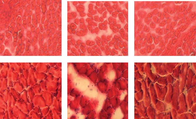
of offspring muscle. There were no obvious adipocytes visible
in muscle cross-section at day 1 (but at day 15,
mature adipocytes were visible in OB offspring SM; metfor-
min inhibited the formation of intramuscular adipocytes in
0 CON OB MET
CON OB MET
CON OB MET
offspring SM induced by the maternal HFD
Maternal metformin administration increased glucose uptake
of offspringThere were no differences in the glucose concentration
Figure 1 AMPK phosphorylation in day-1 and -15 offspring hindleg muscle,and day-60 male offspring gastrocnemius muscle of CON, OB and MET
among treatments at a single time point However,
mothers. Data are expressed as mean±s.e., n ¼ 6. #Po0.05 versus CON;
when the total glucose disposal was compared by combining
(#)Po0.1 versus CON; and *Po0.05 versus OB.
the incremental area under the curve, offspring of OB damhad a lower blood glucose disposal rate compared withthose of CON and MET mothers at both days 15 and 60.
whereas there was no difference between OB and MET
The improvement of glucose uptake following metformin
mothers (Supplementary Figure 2A). Dietary intake did not
administration was associated with enhanced Akt signaling
differ substantially between groups (Supplementary Figure
(Supplementary Figure 3).
2D). OB offspring showed greater weight gain than CONoffspring throughout the experiment whereasMET offspring was intermediate. At day 60, the gonadal fat of
Maternal HFD and metformin administration had no effects
OB male offspring (1.05±0.17 g) was much heavier than that
on muscle fiber type distribution in the offspring SM
of CON offspring (0.57±0.06 g, Po0.05) and tended to be
Myosin heavy-chain isoforms in postnatal mice muscle were
heavier than that of MET offspring (0.74±0.07 g, P ¼ 0.08;
separated (Supplementary Figure 4). There are two develop-
Supplementary Figures 2B and C).
mental myosin heavy-chain isoforms, namely, embryonicand neonatal myosin heavy-chain isoform, present in new-born mouse gastrocnemius, which are replaced by adultisoforms, including type IIA, IIX and IIB by day 14.As
Maternal metformin administration stimulated AMPK activityin offspring SM
shown in Supplementary Figure 4, no difference in muscle
AMPK was activated in the SM of MET offspring. Phosphor-
fiber composition was detected among different treatments
ylation of AMPK at Thr 172 in OB offspring SM was reduced
at all time points. Also, type I fiber was indistinguishable at
compared with CON mothers (Po0.05 at days 15 and 60,
day 60 with this gel electrophoresis method.
and P ¼ 0.06 at day 1, The phosphorylation ofAMPK at Thr 172 was rescued in MET offspring SM at the
Mitochondria DNA copy number and muscle metabolic activity
three time points examined compared with OB offspring SM.
in the offspring was affected by maternal HFD and metformin
Using western blotting, no significant difference in the
protein content of AMPKa subunit was observed (data not
density. Although we found no difference in muscle fiber
Nutrition and Diabetes
Maternal obesity, metformin and offspring muscleJF Tong et al
CON OB MET
CON OB MET
CON OB MET
CON OB MET
CON OB MET
CON OB MET
CON OB MET
CON OB MET
CON OB MET
CON OB MET
CON OB MET
CON OB MET
Figure 2 Contents of b-catenin, myogenic markers and PPAR-g in day-1 and -15 offspring hindleg muscle, and day-60 male offspring gastrocnemius muscleof CON, OB and MET mothers. (a) Beta-catenin content; (b) MyoD content; (c) Myogenin content; (d) PPARgamma content. Data are expressed as mean±s.e.,n ¼ 6. #Po0.05 versus CON; (#)Po0.1 versus CON; *Po0.05 versus OB; and (*)Po0.1 versus OB.
Figure 3 Hematoxylin and eosin (H&E) staining of day-1 and -15 offspring muscle of CON, OB and MET mothers. A section of dorsum of the trunk was dissected,embedded in OCT and frozen in liquid N2. Cryofixed muscle specimens were cross-sectioned at 10-um thickness and stained with H&E, 100 magnification.
(a) CON muscle at day 1; (b) OB muscle at day 1; (c) MET muscle at day 1; (d) CON muscle at day 15; (e) OB muscle at day 15; (f) MET muscle at day 15.
composition among different treatments, mitochondrial
increased mitochondrial copy number in the offspring at day
DNA copy number was decreased in OB offspring at days
15. Moreover, mitochondrial function in offspring SM was
15 and 60 Metformin administration to OB dams
affected by maternal HFD and metformin administration, as
Nutrition and Diabetes
Maternal obesity, metformin and offspring muscle
IAUC glucose
IAUC glucose
Blood glucose (mg/dl)
Time after glucose injection (min)
Figure 4 Glucose tolerance test (GTT) on postnatal day-15 and -60 offspring of CON, OB, and MET mothers. (a) Intraperitoneal GTT in day-15 male offspringmice. The histogram represents the incremental area under the respective glucose curve. (b) Intraperitoneal GTT in postnatal day-60 male offspring mice. Thehistogram represents the incremental area under the glucose curve. Data are expressed as mean±s.e., n ¼ 4 for both ages. #Po0.05 versus CON; (#)Po0.10 versusCON; *Po0.50 versus OB; and (*)Po0.50 versus OB.
mtDNA copy number, citrate synthase and b-HAD activity in day-1
One explanation is that MO programs hypothalamic regula-
and -15 offspring hindleg muscle, and day-60 male offspring gastrocnemius
tion of BW and energy homeostasis.MO in rats does
muscle of CON, OB and MET dams
slightly increase energy intake in offspring.Offspring of
obese rat mothers are hyperphagic.However, in ourstudy, we only observed a trend toward a higher dietary
mtDNA copy numbera
intake in OB offspring compared with CON offspring, which
could be because of our small sample size. Also, the diet used
in our study differed from that in the published rat studies,and offspring may have distinctive preference for sugary and
Citrate synthaseb
fatty items in different diets. In addition, strain differences
have been noted in the response to The observation
that OB offspring showed more weight gain and adipositywhile lacking a significant difference in diet intake suggests
that hyperphagia may not be the only cause of obesity but
differences in basal energy expenditure may also contribute.
SM is the main tissue responsible for glucose and fatty acidoxidation. Reduction in SM oxidative capacity could lead to
Abbreviations: b-HAD, b-hydroxyacyl-CoA dehydrogenase; Cyt C, cytochrome
more offspring energy storage and BW gain. Mitochondria
C oxidase II; mtDNA, mitochondrial DNA; UCP2, uncoupling protein 2.
amtDNA copy number was measured using Cyt C mtDNA gene compared with
are the main sites of nutrient oxidation to generate ATP.
genomic UCP2 gene. bActivity was expressed in mmol per min per g wet
Citrate synthase is a key enzyme in the tricarboxylic acid
weight of muscle. Mean±s.e. *Po0.05 versus CON and #Po0.05 versus OB.
cycle, and b-HAD is a key enzyme in fatty acid oxidation.
Both enzymes are good markers of SM oxidative capacity.Our data showed that both mitochondrial content andfunction were impaired in OB offspring SM, indicating the
evidenced by the decrease of citrate synthase activity and
impairment of SM oxidative capacity. Maternal metformin
b-HAD activity in OB offspring and restoration of their
administration rescued mitochondrial density and function.
activity after maternal metformin administration
In adult muscle, activation of AMPK enhances the oxidativecapacity of SM via a PPAR-g coactivator-1a-mediatedmechanism.Mitochondrial dysfunction leads to obesity,insulin resistance and type 2 diabetThus, our results
support the notion that low oxidative capacity in muscle hasa role in the etiology of obesity.
Both obesity and diabetes are increasing at alarming rates
AMPK has a critical role in maintaining systemic and
and among a younger and younger MO
cellular energy status. The growing appreciation that AMPK
predisposes offspring to obesity and diabetes, which also
coordinates anabolic and catabolic metabolic processes
contributes to the increase in obesity, especially in children.
suggests that it may be an attractive target for preventing
Nutrition and Diabetes
Maternal obesity, metformin and offspring muscleJF Tong et al
diabetes and related metabolic diseases. Physiological activa-
Conflict of interest
tion by exercise or pharmacological activation of AMPKimproves blood glucose disposal, lipid profile and blood
The authors declared no conflict of interest.
pressure in insulin-resistant humans and rodents, whichindicates that AMPK is an effective drug target in thetherapeutic applications in diabetes and other metabolic
Using a sheep MO model, we detected that MOdownregulates AMPK activity in fetal SM.This observation
The research is supported by the National Institute of Child
prompted us to investigate whether activation of AMPK in
Health & Human Development: R01HD067449, and the
fetal and neonatal SM could have a positive effect on its
National Center for Research Resources of NIH and the
development and function. In the present study, we elected
to continue metformin administration throughout lactation
and suckling, as SM development during this period in miceis approximately similar to the development that occurs
from mid to late gestation in sheep and humans. Metforminreadily crosses the Metformin is commonly
1 Petersen KF, Dufour S, Shulman GI. Decreased insulin-stimulated
used to treat type 2 diabetes, and its effect is believed to be
ATP synthesis and phosphate transport in muscle of insulin-resistant offspring of type 2 diabetic parents. PLoS Med 2005; 2:
mainly mediated by AMPK.Our data show that
metformin administration during pregnancy and lactation
2 Galtier F, Raingeard I, Renard E, Boulot P, Bringer J. Optimizing
increased AMPK activity in offspring SM of obese mothers.
the outcome of pregnancy in obese women: from pregestational
b-Catenin is a key mediator of the canonical Wnt/
to long-term management. Diabetes Metab 2008; 34: 19–25.
3 Siega-Riz AM, Siega-Riz AM, Laraia B. The implications of
b-catenin signaling pathway that is essential for myogenesis,
maternal overweight and obesity on the course of pregnancy
and blocking the b-catenin pathway reduces the total
and birth outcomes. Matern Child Health J 2006; 10: 153–156.
number of MyoD and myogenin belong to
4 Morino K, Petersen KF, Dufour S, Befroy D, Frattini J, Shatzkes N
the family of muscle-specific transcription factors that
et al. Reduced mitochondrial density and increased IRS-1 serinephosphorylation in muscle of insulin-resistant offspring of type 2
control SM differentiation;their expression correlates with
diabetic parents. J Clin Invest 2005; 115: 3587–3593.
myogenesis. We observed that b-catenin, MyoD and myo-
5 Fowden AL, Ward JW, Wooding FP, Forhead AJ, Constancia M.
genin were downregulated in fetal OB SM, which is
Programming placental nutrient transport capacity. J Physiol
consistent with our previous observation in sheep
2006; 572: 5–15.
6 Nathanielsz PW. Animal models that elucidate basic principles of
PPAR-g is a critical regulator of adipogenesis. Increased
the developmental origins of adult diseases. Ilar J 2006; 47: 73–82.
expression of PPAR-g leads to differentiation of pluripotent
7 Barker DJ. Fetal programming of coronary heart disease. Trends
cells to Our observation of increased PPAR-g in
Endocrinol Metab 2002; 13: 364–368.
OB offspring SM indicates that adipogenesis was upregu-
8 Armitage JA, Khan IY, Taylor PD, Nathanielsz PW, Poston L.
Developmental programming of the metabolic syndrome by
lated, a conclusion confirmed by the presence of increased
maternal nutritional imbalance: how strong is the evidence from
numbers of intramuscular adipocytes in OB offspring SM,
experimental models in mammals? J Physiol 2004; 561: 355–377.
detected in OB offspring SM. Maternal metformin adminis-
9 Zhu MJ, Han B, Tong J, Ma C, Kimzey JM, Underwood KR et al.
tration reversed these changes, indicating that metformin is
AMP-activated protein kinase signalling pathways are downregulated and skeletal muscle development impaired in fetuses
effective in improving SM development in fetuses and
of obese, over-nourished sheep. J Physiol 2008; 586: 2651–2664.
neonatal mice born to obese mothers. Whether this
10 Tong JF, Yan X, Zhu MJ, Ford SP, Nathanielsz PW, Du M. Maternal
beneficial effect is due to the positive impacts of metformin
obesity downregulates myogenesis and beta-catenin signaling in
on maternal whole-body energy metabolism, improved
fetal skeletal muscle. Am J Physiol Endocrinol Metab 2009; 296:E917–E924.
insulin sensitivity and intrauterine environment or a direct
11 Lowell BB, Shulman GI. Mitochondrial dysfunction and type 2
action on the fetuses of metformin that has crossed the
diabetes. Science 2005; 307: 384–387.
placenta remains to be clarified.
12 Ozanne SE, Jensen CB, Tingey KJ, Storgaard H, Madsbad S, Vaag AA.
Currently, metformin administration is avoided during
Low birthweight is associated with specific changes in muscleinsulin-signalling protein expression. Diabetologia 2005; 48: 547–552.
human pregnancy because of the lack of data about its effects
13 Selak MA, Storey BT, Peterside I, Simmons RA. Impaired oxidative
on fetal development.Our data show that metformin
phosphorylation in skeletal muscle of intrauterine growth-
administration during pregnancy and lactation has no
retarded rats. Am J Physiol Endocrinol Metab 2003; 285: E130–E137.
obvious negative effect on overall fetal and offspring
14 Du M, Yan X, Tong JF, Zhao J, Zhu MJ. Maternal obesity,
inflammation, and fetal skeletal muscle development. Biol Reprod
2010; 82: 4–12.
appeared healthy. In addition, our data clearly show that
15 Mermelstein CS, Portilho DM, Mendes FA, Costa ML, Abreu JG.
metformin administration during pregnancy and lactation
Wnt/beta-catenin pathway activation and myogenic differentia-
improves the development of fetal SM of obese mothers,
tion are induced by cholesterol depletion. Differentiation 2007;75: 184–192.
which may provide a promising strategy to prevent the fetal-
16 Armstrong DD, Esser KA. Wnt/beta-catenin signaling activates
programming effect of obesity during pregnancy on fetal and
growth-control genes during overload-induced skeletal muscle
offspring SM development.
hypertrophy. Am J Physiol 2005; 289: C853–C859.
Nutrition and Diabetes
Maternal obesity, metformin and offspring muscle
17 Cossu G, Borello U. Wnt signaling and the activation of
35 Sparks LM, Xie H, Koza RA, Mynatt R, Hulver MW, Bray GA et al.
myogenesis in mammals. EMBO J 1999; 18: 6867–6872.
A high-fat diet coordinately downregulates genes required for
18 Yamanouchi K, Hosoyama T, Murakami Y, Nishihara M.
mitochondrial oxidative phosphorylation in skeletal muscle.
Myogenic and adipogenic properties of goat skeletal muscle stem
Diabetes 2005; 54: 1926–1933.
cells. J Reprod Dev 2007; 53: 51–58.
36 Call JA, Voelker KA, Wolff AV, McMillan RP, Evans NP, Hulver MW
19 Pan W, Jia Y, Wang J, Tao D, Gan X, Tsiokas L et al. Beta-catenin
et al. Endurance capacity in maturing mdx mice is markedly
regulates myogenesis by relieving I-mfa-mediated suppression of
enhanced by combined voluntary wheel running and green tea
myogenic regulatory factors in P19 cells. Proc Natl Acad Sci USA
extract. J Appl Physiol 2008; 105: 923–932.
2005; 102: 17378–17383.
37 Agbulut O, Noirez P, Beaumont F, Butler-Browne G. Myosin heavy
20 Spiegelman BM, Puigserver P, Wu Z. Regulation of adipogenesis
chain isoforms in postnatal muscle development of mice.
and energy balance by PPARgamma and PGC-1. Int J Obes Relat
Biol Cell 2003; 95: 399–406.
Metab Disord 2000; 24(Suppl 4): S8–S10.
38 Page KC, Malik RE, Ripple JA, Anday EK. Maternal and
21 Aguiari P, Leo S, Zavan B, Vindigni V, Rimessi A, Bianchi K et al.
postweaning diet interaction alters hypothalamic gene expres-
High glucose induces adipogenic differentiation of muscle-
sion and modulates response to a high-fat diet in male offspring.
derived stem cells. Proc Natl Acad Sci USA 2008; 105: 1226–1231.
Am J Physiol Regul Integr Comp Physiol 2009; 297: R1049–R1057.
22 Hardie DG. AMP-activated protein kinase: a key system mediat-
39 Kirk SL, Samuelsson AM, Argenton M, Dhonye H, Kalamatianos T,
ing metabolic responses to exercise. Med Sci Sports Exerc 2004; 36:
Poston L et al. Maternal obesity induced by diet in rats permanently
influences central processes regulating food intake in offspring. PLoS
23 Hardie DG. New roles for the LKB1-AMPK pathway. Curr Opin
One 2009; 4: e5870.
Cell Biol 2005; 17: 167–173.
40 Nivoit P, Morens C, Van Assche FA, Jansen E, Poston L, Remacle C
24 Jakobsen SN, Hardie DG, Morrice N, Tornqvist HE. 50-AMP-
et al. Established diet-induced obesity in female rats leads
activated protein kinase phosphorylates IRS-1 on Ser-789 in
to offspring hyperphagia, adiposity and insulin resistance.
mouse C2C12 myotubes in response to 5-aminoimidazole-4-
Diabetologia 2009; 52: 1133–1142.
carboxamide riboside. J Biol Chem 2001; 276: 46912–46916.
41 Samuelsson AM, Matthews PA, Argenton M, Christie MR,
25 Dagon Y, Avraham Y, Berry EM. AMPK activation regulates
McConnell JM, Jansen EH et al. Diet-induced obesity in female
apoptosis, adipogenesis, and lipolysis by eIF2alpha in adipocytes.
mice leads to offspring hyperphagia, adiposity, hypertension, and
Biochem Biophys Res Commun 2006; 340: 43–47.
insulin resistance: a novel murine model of developmental
26 Habinowski SA, Witters LA. The effects of AICAR on adipocyte
programming. Hypertension 2008; 51: 383–392.
differentiation of 3T3-L1 cells. Biochem Biophys Res Commun 2001;
42 Bruce CR, Anderson MJ, Carey AL, Newman DG, Bonen A,
286: 852–856.
Kriketos AD et al. Muscle oxidative capacity is a better predictor of
27 Giri S, Rattan R, Haq E, Khan M, Yasmin R, Won JS et al. AICAR
insulin sensitivity than lipid status. J Clin Endocrinol Metab 2003;
inhibits adipocyte differentiation in 3T3L1 and restores meta-
88: 5444–5451.
bolic alterations in diet-induced obesity mice model. Nutr Metab
43 Atherton PJ, Babraj J, Smith K, Singh J, Rennie MJ, Wackerhage H.
(Lond) 2006; 3: 31.
Selective activation of AMPK-PGC-1alpha or PKB-TSC2-mTOR
28 Hawley SA, Gadalla AE, Olsen GS, Hardie DG. The antidiabetic
signaling can explain specific adaptive responses to endurance or
drug metformin activates the AMP-activated protein kinase
resistance training-like electrical muscle stimulation. FASEB J
cascade via an adenine nucleotide-independent mechanism.
2005; 19: 786–788.
Diabetes 2002; 51: 2420–2425.
44 Suwa M, Egashira T, Nakano H, Sasaki H, Kumagai S. Metformin
29 Lee JO, Lee SK, Jung JH, Kim JH, You GY, Kim SJ et al. Metformin
increases the PGC-1alpha protein and oxidative enzyme activities
induces Rab4 through AMPK and modulates GLUT4 transloca-
possibly via AMPK phosphorylation in skeletal muscle in vivo.
tion in skeletal muscle cells. J Cell Physiol 2010; 226: 974–981.
J Appl Physiol 2006; 101: 1685–1692.
30 Zou MH, Kirkpatrick SS, Davis BJ, Nelson JS, Wiles IV WG,
45 Hardie DG. Biochemistry. Balancing cellular energy. Science 2007;
Schlattner U et al. Activation of the AMP-activated protein kinase
315: 1671–1672.
by the anti-diabetic drug metformin in vivo. Role of mito-
46 Feig DS. Metformin therapy for gestational diabetes mellitus: are
chondrial reactive nitrogen species. J Biol Chem 2004; 279:
we there yet? Nat Clin Pract Endocrinol Metab 2008; 4: 654–655.
47 Kovo M, Kogman N, Ovadia O, Nakash I, Golan A, Hoffman A.
31 Ma TC, Buescher JL, Oatis B, Funk JA, Nash AJ, Carrier RL et al.
Carrier-mediated transport of metformin across the human
Metformin therapy in a transgenic mouse model of Huntington's
placenta determined by using the ex vivo perfusion of the
disease. Neurosci Lett 2007; 411: 98–103.
placental cotyledon model. Prenat Diagn 2008; 28: 544–548.
32 Folch J, Lees M, Sloane Stanley GH. A simple method for the
48 Paglia MJ, Coustan DR. The use of oral antidiabetic medications
isolation and purification of total lipids from animal tissues. J Biol
in gestational diabetes mellitus. Curr Diab Rep 2009; 9: 287–290.
Chem 1957; 226: 497–509.
49 Sabourin LA, Rudnicki MA. The molecular regulation of myogen-
33 Underwood KR, Tong J, Zhu MJ, Shen QW, Means WJ, Ford SP
esis. Clin Genet 2000; 57: 16–25.
et al. Relationship between kinase phosphorylation, muscle fibertyping, and glycogen accumulation in longissimus muscle of beefcattle with high and low intramuscular fat. J Agric Food Chem
This work is licensed under the Creative
2007; 55: 9698–9703.
34 Guo W, Jiang L, Bhasin S, Khan SM, Swerdlow RH. DNA
Alike 3.0 Unported License. To view a copy of this
mtDNA copy number determination. Mitochondrion 2009; 9:
Supplementary Information accompanies the paper on the Nutrition and Diabetes website (http://www.nature.com/nutd)
Nutrition and Diabetes
Source: http://vuir.vu.edu.au/30770/1/Metformin%20mitigates%20the%20impaired%20development%20of%20skeletal%20muscle%20in%20the%20offspring%20of%20obese%20mice.pdf
Life Science Trends 2014 Feature Article: Big Data in the Life Sciences D. Alexander, C. Hamilton, A. McDowell, B. McMerty, N. Burns, C. Hancock, J. McLaughlin About Life Science Trends 2014 About this Report Each year, Carlyle Conlan provides an overview of trends and innovations in the life science
Three reasons not to buy the NicoTestTM genetic test GeneWatch UK What is the NicoTestTM? NicoTest is a new genetic test kit being marketed directly to the public via the internet(www.nicotest.com). It is marketed by a company called g-Nostics Ltd which hopes tosell it more widely in the future, both "over the counter" and via doctors in the NationalHealth Service. G-Nostics is a "spin out" company from Oxford University. The universityis a shareholder in the company and the test is based on research by Dr Robert Waltonin the university's Department of Clinical Pharmacology1. Dr Walton is Chief ScientificOfficer, Lead Inventor and co-founder of the company2.